


Rectus Femoris: Comprehensive Guide to Anatomy & Function
The Anatomy, Biomechanics, and Pathology of the Rectus Femoris
The human lower limb is a masterpiece of evolutionary engineering, designed to balance the competing demands of structural stability and dynamic mobility. At the center of this functional dichotomy sits the quadriceps femoris, a massive muscle group occupying the anterior compartment of the thigh. While three members of this group—the vastus lateralis, vastus medialis, and vastus intermedius—are monoarticular muscles crossing only the knee joint, the fourth member stands apart. This unique muscle is the rectus femoris.
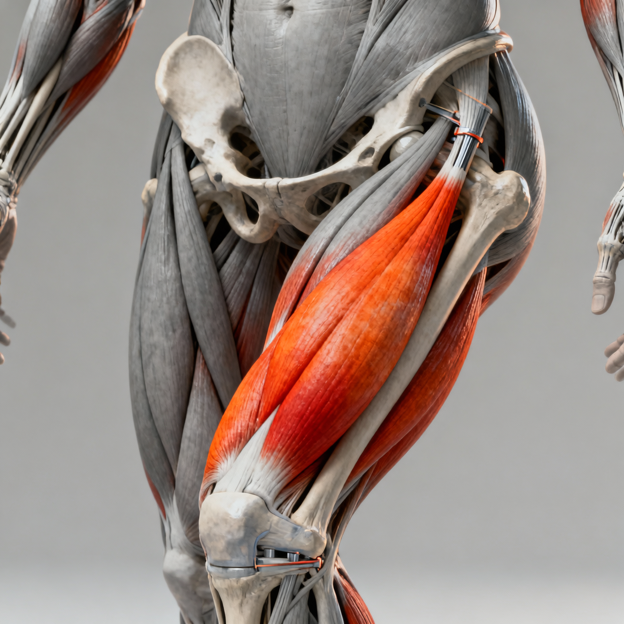
Characterized by its straight path down the center of the thigh and its bipennate fiber arrangement, this muscle is the only superficial component of the quadriceps that crosses two major joints: the hip and the knee. This dual-joint architecture endows it with complex biomechanical responsibilities, making it indispensable for actions ranging from simple walking to explosive jumping and sprinting. Simultaneously, this multi-joint involvement subjects the muscle to unique mechanical stresses, rendering it highly susceptible to injuries in athletic populations.
Anatomical Profile and Structural Overview
The name of the muscle is derived directly from its physical characteristics: “rectus” translates from Latin as “straight,” while “femoris” refers to the femur or thigh. True to its name, it descends almost vertically along the anterior midline of the thigh, situated superficially to the vastus intermedius and flanked by the vastus medialis and vastus lateralis.
Origins and Proximal Attachment
Unlike its fellow quadriceps muscles, which arise from the shaft of the femur, the proximal attachments of this muscle anchor it directly to the pelvis. This pelvic connection is accomplished via two distinct, robust tendinous heads:
- The Straight Head (Direct Head): Arises from the anterior inferior iliac spine (AIIS), a bony projection on the anterior aspect of the ilium.
- The Reflected Head (Indirect Head): Originates from a groove or sulcus located just superior to the acetabular rim (the hip socket) and the capsule of the hip joint itself.
Some anatomical studies also identify a third, deep recurrent head that blends with the iliofemoral ligament. These proximal tendons merge shortly after their origin to form a unified aponeurosis, which transitions into a broad, fleshy muscle belly.
Distal Insertion and the Quadriceps Tendon
As the muscle belly descends, its fibers exhibit a classic bipennate arrangement. The muscle fibers run obliquely toward a central tendon, resembling the barbs of a feather. This structural design maximizes the physiological cross-sectional area, allowing the muscle to pack a high density of contractile units into a relatively narrow pathway.
In the distal third of the thigh, the muscle belly transitions into a flat, thick tendon. This tendon converges with the tendons of the three vastus muscles to form the powerful quadriceps tendon. The superficial fibers of the rectus tendon pass directly over the anterior surface of the patella (kneecap) to insert into the tibial tuberosity via the patellar ligament. This complex arrangement means that any force generated by the muscle is transmitted directly through the patellar lever arm, facilitating efficient movement.
The Biological Profile of the Rectus Femoris
To understand its physical place in the body, it is helpful to look at its direct biological profile, mapping out its neurological, vascular, and structural details.
| Biological Attribute | Anatomical Specification |
| Muscle Classification | Bipennate, fusiform, biarticular (dual-joint) muscle |
| Primary Actions | Knee extension (straightening) and hip flexion (lifting the thigh) |
| Proximal Origin | AIIS (straight head) and superior acetabular groove (reflected head) |
| Distal Insertion | Base of the patella, continuing via patellar ligament to tibial tuberosity |
| Innervation | Femoral nerve (originating from spinal levels L2, L3, and L4) |
| Vascular Supply | Descending branch of the lateral circumflex femoral artery |
| Antagonists | Hamstrings (biceps femoris, semitendinosus, semimembranosus) |
| Synergists (Hip) | Iliopsoas, tensor fasciae latae, sartorius, pectineus |
| Synergists (Knee) | Vastus lateralis, vastus medialis, vastus intermedius |
This structural blueprint allows the muscle to act as a bridge between the pelvic girdle and the lower leg, coordinating complex movement patterns across the entire lower extremity.
Biomechanical Mechanics and Joint Coordination
Because it crosses both the hip and knee joints, the biomechanical contributions of this muscle are highly complex and dependent on the relative positions of both joints. This dual-joint functionality introduces a phenomenon known as the length-tension relationship, which heavily influences how the muscle produces force.
                  [ Pelvis (Origin: AIIS) ]
                             |
                             |  (Crosses Hip Joint)
                             v
               [ Rectus Femoris Muscle Belly ]
                             |
                             |  (Crosses Knee Joint)
                             v
                 [ Patella / Tibial Tuberosity ]
1. Knee Extension
When the hip is held in a fixed position, contracting the muscle pulls the patella superiorly. This action pulls on the patellar ligament, rotating the tibia forward around the femoral condyles and extending the knee. While the vastus muscles are responsible for the vast majority of knee extension torque, the rectus femoris provides crucial dynamic stabilization, especially during high-velocity movements.
2. Hip Flexion
When the knee is stabilized or held in flexion, contraction of the muscle pulls the femur toward the anterior pelvis, flexing the hip. It works alongside primary hip flexors like the iliopsoas. This is highly apparent during the swing phase of gait, where the leg must be lifted and swung forward to take a step.
3. The Challenge of Active and Passive Insufficiency
The biarticular nature of the rectus femoris exposes it to two functional limitations that govern its performance:
- Active Insufficiency: This occurs when a multi-joint muscle is shortened across both joints simultaneously, reducing its ability to generate active tension. For example, if you sit with your hip fully flexed and attempt to fully extend your knee, you will find the extension movement feels weak and difficult. The rectus femoris is already highly shortened at the hip, leaving its sarcomeres with minimal overlap to produce further contraction torque at the knee.
- Passive Insufficiency: This occurs when a multi-joint muscle is fully elongated across both joints simultaneously, reaching the limit of its passive stretch. If you extend your hip backward while flexing your knee toward your glutes, you will feel a dramatic, tight stretch along the front of your thigh. This passive tension can restrict the range of motion of the joints if the muscle is excessively tight.
4. Energy Transfer and the “Catapult” Action
During dynamic activities like kicking a soccer ball or sprinting, the rectus femoris acts as an energy-transfer conduit. During the backswing of a kick, the hip is extended and the knee is flexed. This stretches the rectus femoris across both joints, storing elastic energy within its collagen-rich tendinous tissues.
As the forward swing begins, the hip flexes rapidly while the knee temporarily remains flexed. This action releases the stored elastic energy like a catapult, driving high-velocity knee extension just before foot-to-ball contact. Without this coordinated biarticular action, human movement would lose much of its explosive power.
Pathophysiology and Common Clinical Conditions
Given its superficial location, high percentage of fast-twitch muscle fibers, and dual-joint architecture, this muscle is one of the most frequently injured structures in the lower body. It is particularly vulnerable in sports that require sudden acceleration, jumping, or repetitive kicking, such as soccer, rugby, football, and track and field.
Rectus Femoris Strains
Muscle strains typically occur during eccentric contractions, where the muscle is forced to lengthen while actively trying to contract. This happens during the transition phase of sprinting or the terminal backswing of a kick. Strains are classified into three distinct grades:
- Grade I (Mild): Microscopic tearing of muscle fibers. The patient experiences localized tenderness and minimal swelling, with no significant loss of strength.
- Grade II (Moderate): Partial tearing of the muscle fibers. This presents with distinct pain, visible swelling, localized bruising, and a noticeable reduction in both hip flexion and knee extension strength.
- Grade III (Severe): Complete rupture of the muscle belly or tendon. This is accompanied by sudden, excruciating pain, a popping sensation, extensive hematoma formation, and a palpable defect or “gap” in the anterior thigh.
Distal and Proximal Tendinopathy
Repetitive load without adequate recovery can lead to microtrauma at the tendinous attachments, resulting in tendinopathy. Proximal tendinopathy often presents as deep, aching groin pain near the AIIS, which is easily aggravated by high-intensity sprinting or hip flexion. Distal tendinopathy manifests as anterior knee pain just above the patella, often referred to as quadriceps tendinitis.
Deep Muscle Hematomas and Myositis Ossificans
Because of its superficial position on the front of the thigh, the rectus femoris is highly susceptible to direct impacts, such as a blunt blow from an opponent’s knee during contact sports (commonly known as a “charley horse”). If a deep muscle contusion is treated too aggressively with deep tissue massage, premature heat application, or forced stretching during the acute healing phase, a condition called myositis ossificans can develop.
In this condition, calcifications form within the muscle tissue, as the body mistakenly deposits calcium and bone-like structures inside the healing hematoma. This leads to chronic pain, permanent loss of flexibility, and a prolonged recovery timeline.
Rehabilitation, Strengthening, and Injury Prevention
Preventing injuries or recovering from a strain requires a targeted approach that respects the dual-joint nature of the rectus femoris. Standard single-joint exercises are rarely sufficient to build the required tissue resilience.
Targeted Strengthening Exercises
To build robust muscle fibers capable of withstanding high eccentric loads, exercises must challenge the muscle across its entire functional length.
- The Spanish Squat: This variation utilizes a heavy band anchored behind the knees. By leaning back against the band while maintaining a relatively vertical torso, the vasti and rectus muscles are subjected to intense, continuous isometric tension, which is highly effective for tendon remodeling.
- Reverse Nordic Curls: This bodyweight exercise involves kneeling on the floor with a straight torso and slowly leaning backward from the knees. This movement subjects the rectus femoris to an intense eccentric load while in a position of hip extension, training the muscle to safely handle high tension while elongated.
- Decline Squats: Performing squats on a 15-to-20-degree decline board shifts the center of mass, placing a higher mechanical load on the anterior quadriceps structures.
Mobility and Flexibility
To combat passive insufficiency and maintain pelvic alignment, systematic stretching is essential.
- The Couch Stretch: By placing the knee of the rear leg flat against a wall or couch while stepping the opposite leg forward into a lunge, the hip is extended while the knee is deeply flexed. This directly targets the rectus femoris, relieving tension on the anterior pelvis.
- Active Isolated Stretching: Performing gentle, active stretches of the hamstrings forces the anterior quadriceps to relax via reciprocal inhibition, facilitating safer and deeper mobility work for the anterior thigh.
Exploring Key Questions
How can I tell the difference between a rectus femoris strain and a hip flexor strain?
Because the rectus femoris acts as a hip flexor, distinguishing between a strain of this muscle and other hip flexors (like the iliopsoas) can be difficult. A key diagnostic indicator is knee flexion. If you lie on your stomach and attempt to pull your heel toward your glutes, sharp pain in the front of your thigh indicates a rectus femoris issue.
Because the iliopsoas does not cross the knee joint, bending the knee does not put it under tension. Furthermore, a rectus femoris strain will typically cause localized tenderness directly on the anterior thigh or just below the pelvic bone, whereas iliopsoas discomfort is felt deeper within the groin area.
Why is this muscle more prone to injury than the other three quadriceps muscles?
The primary reason is its biarticular (dual-joint) structure. Crossing two joints means it can be stretched to its limit at both ends simultaneously—such as during the backswing of a kick when the hip is extended and the knee is flexed.
Additionally, it contains a high percentage of type II (fast-twitch) muscle fibers, which are designed for rapid, powerful contractions but fatigue quickly and are more susceptible to structural damage under high eccentric loads.
Can a tight rectus femoris cause lower back pain?
Yes. Because the muscle originates at the anterior inferior iliac spine (AIIS) of the pelvis, chronic tightness or shortening of this muscle pulls the anterior pelvis downward and forward. This causes an anterior pelvic tilt.
An anterior pelvic tilt increases the arch of the lumbar spine (hyperlordosis), compressing the facet joints of the lower back and putting excess strain on the surrounding spinal muscles, which frequently leads to chronic lower back discomfort.
What is the typical recovery time for a moderate rectus femoris strain?
A moderate (Grade II) strain involves partial tearing of the muscle fibers and generally requires four to eight weeks of structured rehabilitation. The recovery process must progress through initial pain management and swelling reduction, followed by sub-maximal isometric strengthening, gradual eccentric loading, and finally, sports-specific plyometrics and speed work. Rushing back to play before restoring full eccentric strength and flexibility carries a high risk of reinjury.
Summary of Functional Dynamics
Ultimately, the rectus femoris is far more than just a muscle on the front of the thigh; it is a critical mechanical bridge connecting the pelvis to the lower leg. Its unique dual-joint architecture allows us to run, jump, kick, and stabilize our lower body with remarkable efficiency. By understanding its complex biomechanics, taking care of its flexibility, and training it through its full length, we can optimize physical performance, maintain healthy pelvic posture, and protect our lower limbs from injury.
Read more:https://articlebytes.co.uk/golden-bachelor-spoilers/